image_pdfimage_print

Opioid-Induced Hyperalgesia (OIH). Opioid medications can worsen your chronic pain.

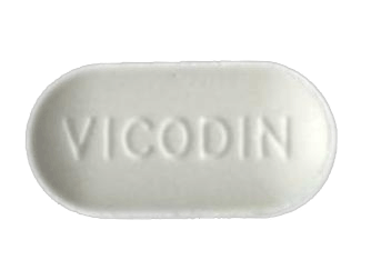
Opioids reduce the intensity of pain signals reaching the brain and diminish the emotional response to pain. Opioids include: Hydrocodone (Vicodin), Codeine, Fentanyl, Hydromorphone, Meperidine, Morphine, Sufentanil, Oxycodone, Oxymorphone, Buprenorphine, Methadone, and Tramadol.

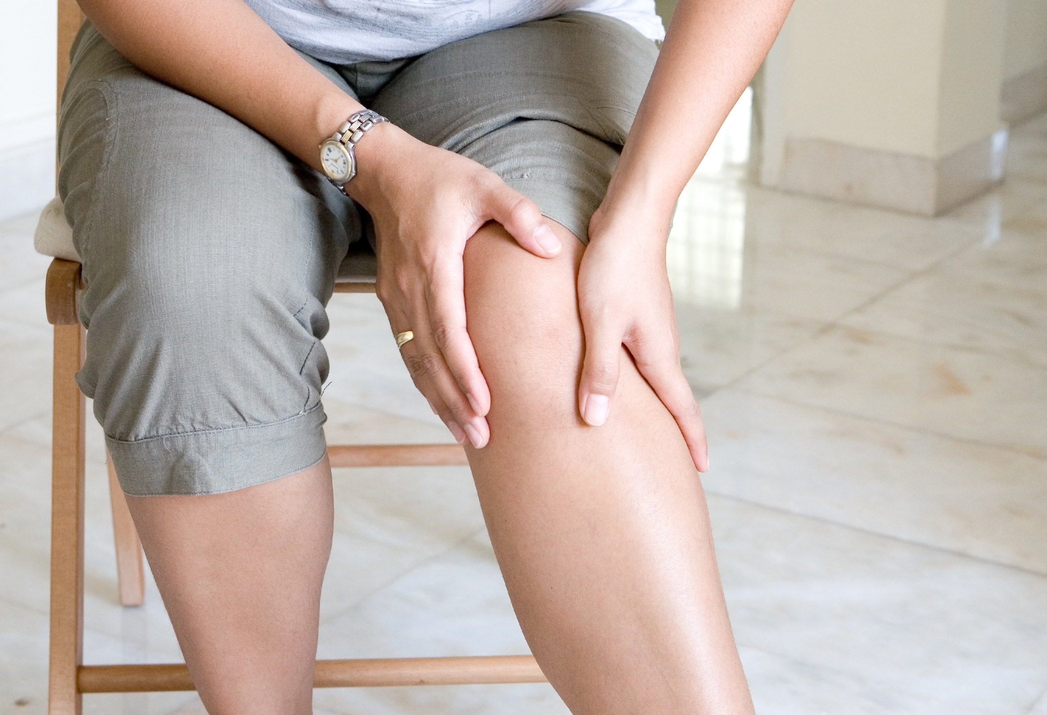
Opioid-induced hyperalgesia (OIH) is a condition whereby opioid therapy has a reverse effect on patients and causes increased sensitivity to certain types of pain. The symptoms of OIH are often difficult to diagnose and symptoms present as a worsening of the patient’s chronic pain with regions of pain spreading to other areas of the body. This worsening pain condition can be incorrectly interpreted as a patient developing a tolerance to the opioid that reduces the drug’s effect. This may not be the case. Prescribing higher dosages of opioids can paradoxically worsen a patient’s pain condition.

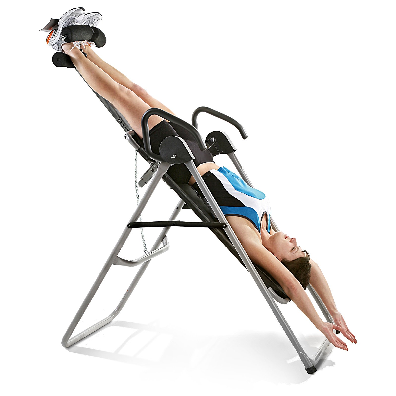
There is no question that getting off opioids can be very difficult and some physicians don’t want to assume the responsibility and risk that can arise from withdrawal and detox. An integrative medical approach is increasingly being sought after by both physician and patient to reduce dependence on opioids and to develop a safer pain management plan.

Acupuncture has been used to treat both chemical addiction and chronic pain. Acupuncture observes the improvement in pain reduction as a reduction in the frequency, intensity, and location of pain. Ideally, progress in the treatment of pain with acupuncture should result in the complete opposite effect of OIH—a reducing and localizing of pain, a normalizing of pain tolerance, and a restoration of body functions (normal bowl movements, proper digestion and stomach emptying, and sustainable energy with better sleep).

It is important for patients to express their concerns with long-term use of opioids with their physician and to establish an exit strategy to get off pain medications before even starting them.

Aging: Why do I hurt?

As a specialty practitioner in pain mitigation, I frequently get asked why people experience an increasing amount of joint and muscle pain with age. Of course, there are many possible answers for this: arthritis, degenerative spinal disc and joint conditions, neuropathic conditions, dietary inflammatory agents, etc. However, the majority of generalized joint and muscle pain are rooted in hypokinetic disease with disuse atrophy.

The terms “hypokinetic disease” and “disuse atrophy” sound like serious medical conditions, but what they essentially refer to is muscle breakdown due to decreased physical activity. When our activity levels significantly drop off our muscles begin to shrink and eventual breakdown of the actual muscle fibers. This process can result in increased sensitivity of the nerves, inflammation, and joint instability.

Acupuncture, deep tissue massage, electro-stimulation, and non-steroidal injection therapy are effective means for eliminating muscle soreness, treating trigger points and hypersensitive nerves, as well as improving circulation and healthy muscle tension.

The number one self-help fix is of course increasing the level of physical activity and re-engaging the muscles. The old myths of having to put in an hour at the gym 3-4 days per week to maintain physical conditioning have long been dispelled. Research has shown that strength exercises performed only once per week can maintain muscles strength in many muscle groups. Now I’m not advocating only exercising once per week; I’m just attempting to reduce the opportunity for excuse making. So consider that while you are not exercising because you hurt, you may be hurting because you are not exercising.

Cupping on Elite Athletes

Cupping is a good tool for Sports Medicine; however, cupping may not be a good idea for an elite athlete prior to an event.

Traditional Method of CuppingCupping (vacuum technique)

Cupping causes several physiological events to happen. First, the vacuum pressure from the cups pulls in blood and interstitial fluid causing hyperemia (increased blood flow) along with skin stretching and opening of the pores.

Second, the pressure in the small capillary beds for blood within the dermis rupture and hemorrhage to form a hematoma (bruise).

img_2076img_2075
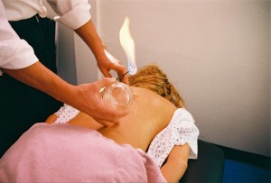
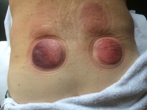
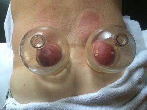
Third, the capillary beds then undergo hemostasis (clotting) to stop the leaking of blood into the interstitial spaces.

Forth, an inflammatory response occurs to remove damaged tissue and mobilize 0405
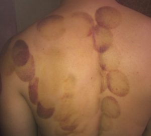
cellular proliferation to the site.

Fifth, tissue repair begins along with angiogenesis (formation of new blood vessels).

Now that you know what cupping does, why would you intentionally break down blood vessels, create inflammation, increase opportunity for fatigue, and compromise the skin’s shielding effect immediately before a world championship competition when winning and losing is measured within a one-thenth of a second?

In my opinion, this specific case of cupping in sports is for a psychological advantage and not a physiological one. If an athlete thinks cupping or Kinesio tape will give them an edge then maybe it will, even if it is just a boost in their confidence.

This confidence may well be nullified by an equal boost in the confidence of competitors by suggesting a weakness or injury.

Inversion Tables for Lumbar Discogenic Pain

WESLO-Weslo-Inversion-Flex-System-Inversion-Table_1_0What is Discogenic Pain?

Discogenic pain presents as localized pain at the spine, most often the lumbar spine that worsens with lifting, coughing, sneezing, or straining during a bowel movement. Discogenic pathologies include bulging or herniated intervertebral discs, degeneration of a disc, dehydration of the disc nucleus, or tears in the fibrous capsule (annulus) of the disc. A malformed or degenerated disc can result in the boney vertebrae moving closer together causing peripheral nerve impingement at the neural foramina.

What do Inversion Tables do?06_Vertebral_and_Intervertebral_Disc_Anatomy

Inversion tables use gravity and your body weight to traction the spine. As the spine stretches, it creates a negative pressure within the intervertebral discs. The negative pressure, in conjunction with osmosis, pulls water into the gelatinous center of the disc called the nucleus pulposus. Stretching and hydration thicken the discs and lengthens the spine. As the discs thicken the separate the vertebra and increase the free space around the nerves as they leave the spine- the result is a healthier and happier you.

What are the contraindications for inversion tables?

Inversion tables take a little coordination and possibly even some assistance. Most inversion tables work by setting the maximum decline angle, locking your ankles into the suspension, then leaning back and slowly tilting backward until your head is below your ankles. This seemingly simple process can be problematic if you have the following issues:

  • Retinal detachment
  • Stroke
  • Recent head injury
  • Spinal injury
  • Bone weakness
  • Lumbar fusions, either surgical or from bone remodeling
  • Severe osteoarthritis of the facet joints of the spine
  • Tears in the vertebral discs
  • Vestibular problems resulting in dizziness and nausea

Geriatric patients or patients with disabilities should not use inversion tables without assistance and clearance from their primary care physician.

When will I feel better?

When patients first begin stretching their spine, it can result in increased discomfort for 1-2 weeks. The reason for this is that the musculature of the spine has contracted to protect and stabilize the injured area. The problem with this is that the increased muscle contraction also creates more pressure on the discs, flattening them out and reducing their ability to rehydrate fully. Increased disc compression with dehydration equals prema ture disc degeneration.

Relief can be immediate but short-lasting. Long-term results will take 4-8 weeks of tractioning five times per week. After the discogenic pain has resolved, a maintenance tractioning program of 3-4 times per week is recommended.

When should I do traction and for how long?

Traction should be performed just before bedtime for 5-10 minutes. Tractioning just before bedtime allows you to lay flat after stretching the discs, so gravity doesn’t recompress the spine before they get a chance to rehydrate.

Stop traction if your pain significantly increases.

Even though your might get slightly worse before getting better, a dramatic worsening in your condition the following traction may indicate that there is a more significant spinal pathology occurring and further diagnosis is required.

Treatment of Plantar Fasciitis Foot Pain

Foot pain is a very common ailment that can range from mild discomfort too a debilitating condition that limits ones quality of life. Plantar fasciitis is a frequently diagnosed condition characterized by chronic heel pain and pain along the sole of the foot. Although there are several conditions that exhibit this pattern, plantar fasciitis displays a symptom called “poststatic dyskinesia,” which is pain with increased intensity when rising from a resting position. People with plantar fasciitis frequently have severe foot pain when they first get out of bed in the morning, but the pain diminishes with activity and later returns as the day wears on.

Multiple Causes of Foot Pain

Plantar fasciitis can have several different causes but is usually a result of repetitive stress injuries to the feet. This condition is commonly seen in patients who stand on hard flat surfaces for prolonged periods of time. Runners and joggers that train on pavement are prone to this condition. Plantar fasciitis can also occur for no known cause or as part of the aging process. A professional medical diagnosis should be made to determine the appropriate treatment approach. In addition to plantar fasciitis, there are several other possibilities that can cause foot pain and require a differential diagnosis, some of these include peroneal tendonitis, gastro-soleus complex, plantar calcaneal bursitis, metatarsalgia, heel fat pad syndrome, and several others.

Scar Tissue and “Bone Spur” Formation

Scar tissue may form at the heel of the foot as a result of chronic irritation and inflammation of the plantar fascia. If the inflammation, scarring, and irritation become chronic they can resulting calcification of the fascia at its attachment point on the calcaneal (heel) bone. This calcified fascia appears on a foot x-ray as an hook shaped bone spur on the sole of the foot at the heel. When fasciitis goes untreated and becomes chronic to the point of bone spur formation, non-invasive techniques may not be effective and surgery is often required.

Treatment Approaches Provided by Medical Practitioners

Practitioners such as chiropractors, physical therapists, acupuncturists, and massage therapists can provide a variety of effective modalities for the treatment of plantar fasciitis. Techniques such as micro current, ultrasound, interferential, acupuncture, transdermal medications, and manual therapies can provide effective and permanent pain relief. Physicians can inject a steroid into fascia attachment at the heel to reduce local inflammation.

Foot Orthotics and Shoe Inserts

The opinion of using inserts and orthotics is more a matter of philosophy than medical science. Some practitioners believe that providing arch support in the form of orthotics will take pressure off the plantar fascia and allow it to heal. Others believe that the arch naturally falls with normal wear and aging of the feet. As the arch begins to break down and drop, there is increased tension on the fascia as the foot elongates, which causes inflammation and pain in the sole of the foot. In this case, practitioners encourage stretching the fascia to allow for the new foot position so the fascia can heal on its own. Hard orthotic inserts are believed by some to only further weaken the arch and prolong irritation of the plantar fascia.

Self-Help Remedies

The #1 most common self-help treatment for plantar fasciitis is golf ball massage. Yep, a plain old golf ball. Placing a golf ball on the ground and then rolling it under the sole of your foot will stretch the fascia and improve circulation. The golf ball is just the right size and shape to effectively massage the sole of the foot.

Heel lift inserts, or just wearing shoes with ½ inch to 1 inch heels, can be beneficial and provide relief for some people when tight calf muscles and/or Achilles tendinitis may be contributing to the foot pain.

Calf stretches with full extension of the toes (pulling the toes up) is a good stretch for the plantar fascia. There are foot supports that can be worn at night that keep the ankle flexed and toes extended to stretch the fascia while you sleep.

Hot water foot soaks. Although ice may feel good and temporarily numb the area, ice causes constriction of blood vessels and slower healing times. Unless there are acute signs of redness, swelling, heat, and pain, or you just need some immediate pain relief, it is best to avoid ice and stick with heat. When in doubt as to use heat or ice, do what provides the most relief.

Tips for reducing joint pain and increasing movement

knee_pain

  • Start moving. Joints are designed to move and reducing movement also reduces synovial fluid lubrication and blood circulation within joints.
  • Perform static flexion of joints for a minimum of 2-3 minutes prior to exercise or prolonged activity. Full flexion of a joint decreases joint pressure and distributes lubricants across the joint space.
  • Move joints through a full range of motion. Limiting joint movement causes uneven wear on joints.
  • Joints that “give out” are typically the result of issues with the muscles and ligaments that cross the joints. Maintaining good muscle tone is essential for joint stability.
  • Edema and swelling in and around joints (joint effusion) can contribute to joint pain. Wearing thin, flexible compression sleeves over joints (i.e. ankles, wrists, knees, and elbows) combined with the “pumping” action of joint movement can reduce or prevent joint swelling.

 Dietary Recommendations

  • Glucosamine sulfate, a supplement found in the exoskeletons of crustaceans, has been shown to ease joint pain and stiffness in joints if taken during early onset. Dose: 1,500 mg (1.5 gm) per day.
  • Omega-3 fatty acids have been shown to have anti-inflammatory and pro-resolution properties. These fatty acids stimulate the clearance of inflammatory debris, promote mucosal antimicrobial defense, protect organs from collateral damage, and enable inflamed tissues to return to homeostasis. Dose: 1,240 mg (1.24 gm) per day.
  • Herbs such as turmeric, ginger, and boswellia may also be beneficial as natural anti-inflammatories. Bromelain, a pineapple enzyme, has also been used as an anti-inflammatory when taken in doses of 200-400 mg per day on an empty stomach.
  • S-adenosylmethionine (SAMe) is found naturally in the body. SAMe supplementation has been found to be as effective as and safer than non-steroidal anti-inflammatory drugs (NSAIDs) but has a slower onset time. Dose: 400 mg 3-4 times per day for 1-12 weeks.